Plant tissue, Animal tissue, Collenchyma, Sclerenchyma and much more……. Class 9 Science Notes Tissue
tissues are the groups of similar cells, having the same origin and performing a specific function. The name tissue was given by Bichat. Tissue first evolved in coelenterates.
- Organs are group of different types of tissues performing some specific functions. Such as Stomach, Heart, etc.
Organ system: The entire organ that comes together to perform some specific functions.
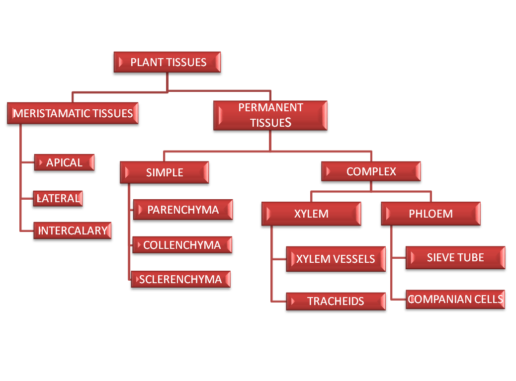
Plant Tissues
A group of cells having same origin and co-operative with one another to performing a similar function is called as a tissue. The study of tissue is called histology. Depending upon constitution of cells, the tissues are of two types:
- Simple tissues – it is made up of similar cells.
- Complex tissues – it is made up of two or more than two types of cells.
Based on the capacity to divide the plant tissues have been classified into two fundamental types:
Meristematic tissue
It is a simple tissue that composed of a group of similar and immature cells which can divide and form new cells.
Characteristics
- Ability to grow and divide
- Absence of intercellular spaces
- Dense cytoplasm
- Mitochondria have simple structure
- Rate of respiration is very high
- Walls are thin, and made of cellulose
The meristematic cells from which all other types of cell are formed depending upon their origin, meristem is of two types: Primary & Secondary.
- Primary meristems: these are those meristematic tissues which are derived directly from the meristem of the embryo. Depending on their position they are of three types:
- Apical meristem: these are present on the tips of the stem, roots and their branches. They produce growth in length.
- Intercalary meristem: These meristematic tissues present at the internodes of the stems and at the leaf-bases. These help in elongation of organs.
- Lateral meristem: The meristem occurs on the sides and takes part in increasing the girth of the plant. This present below the bark and in the vascular bundles in dicot stem of gymnosperm. It is responsible for increasing the diameter of the stem e.g. Cambium.
- Secondary meristems: Meristem is formed secondarily from the permanent tissues. Secondary meristem is usually lateral. They are cylindrical meristem these meristem give rise to secondary tissue that constituent secondary growth. Common examples are Vascular cambium of root, Inter-fascicular vascular cambium of stem, Cork cambium, Wound cambium and Accessory cambia of monocots (yucca).
PERMANENT TISSUE
They are those tissues which have lost capacity to divide and have attained a permanent shape, size and function. The permanent cells may be dead or living. Depend upon the structure; the permanent tissues are classified into the following two types:
Simple permanent tissues
Permanent tissues having all cells Similar in structure and function are called simple tissues. They include protective and supportive tissues.
- Protective tissues: these include epidermis and cork cambium.
- Supporting tissues : these are categorised into three types
Parenchyma
Parenchyma forms the major component within organs. The cells of the parenchyma are generally isodiametric.
- They may be spherical, oval, round, polygonal or elongated in shape.
- Their walls are thin and made up of cellulose. They may either be closely packed or have small intercellular spaces. The parenchyma performs various functions like photosynthesis, storage, secretion.
- They form the ground tissue in the non – woody or soft areas of stems, leaves, roots, flowers, fruits etc.
DIFFERENT TYPES OF PARENCHYMA
Functions
- It is basically for the storage of food e.g. starch
- It also provides turgidity to plants.
- It provides rigidity to tissues.
- Photosynthesis in the form of chlorenchyma.
- Providing buoyancy and storage of metabolic gases in the form of aerenchyma.
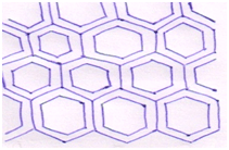
Collenchyma
Collenchyma is made up of living cells which are usually elongated with thick corners. There is absence of intercellular spaces in this tissue. Few chloroplasts are present in the cell of Collenchyma. The Collenchyma occurs in layers below the epidermis in dicotyledonous plants. It consists of cells which are much thickened at the corners due to a deposition of cellulose, hemicellulose and pectin. These cells assimilate food when they contain chloroplasts. Inter cellular spaces are absent. They provide mechanical support to the growing parts of the plant such as young stem and petiole of a leaf.
DIFFERENT TYPES OF COLLENCHYMA
Functions
- It provides mechanical strength to young dicot stem, leaves etc.
- It also provides flexibility to the organ and allows their bending.
- It prevent tearing of leafs.
Sclerenchyma
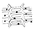
Sclerenchyma consists of long, narrow cells with thick and lignified cell walls having a few or numerous pits. They are usually dead and without protoplasts. On the basis of variation in form, structure, origin and development, Sclerenchyma may be either fibres or sclereids. The fibres are thick-walled, elongated and pointed cells, generally occurring in groups, in various parts of the plant. The sclereids are spherical, oval or Cylindrical, highly thickened dead cells with very.
T.S. OF FIBRES
Functions
- It provides rigidity to leaves, and prevents collapsing during temporary wilting.
- A number of fibre commercially exploited, e.g. jute.
- Sclerenchyma is the chief mechanical tissues of the mature plant organ.
Complex PERMANENT TISSUES
The complex tissues are made of more than one type of cells and these works together as a unit. Xylem and phloem constitute the complex tissues in plants.
Xylem functions as a conducting tissue for water and minerals from roots to the stem and leaves. It also provides mechanical strength to the plant parts.
It is composed of four different kinds of elements, namely, tracheids, vessels, xylem fibres and xylem parenchyma. Gymnosperms lack vessels in their xylem.
- Tracheids are elongated or tube like cells with thick and lignified walls and tapering ends. These are dead and are without protoplasm. In flowering plants, tracheids and vessels are the main water transporting elements.
- Vessel is a long cylindrical tube-like structure made up of many cells called vessel members, each with lignified walls and a large central cavity. The vessel cells are also devoid of protoplasm. The presence of vessels is a characteristic feature of angiosperms.
- Xylem fibres have highly thickened walls and have central lumens. These may either be septate or aseptate. Xylem parenchyma cells are living and thin-walled, and their cell walls are made up of cellulose. They store food materials in the form of starch or fat, and other substances like tannins. The conduction of water takes place by parenchymatous cells
Phloem transports food materials, usually from leaves to other parts of the plant.
Phloem in angiosperms is composed of sieve tube elements, companion cells, and phloem parenchyma and phloem fibres.
Gymnosperms have albuminous cells and sieve cells. They lack sieve tubes and companion cells.
- Sieve tubes elements are also long, tube-like structures arranged longitudinally and are associated with the companion cells. The functions of sieve tubes are controlled by the nucleus of companion cells.
- The companion cells are specialised parenchymatous cells, which are closely associated with sieve tube elements.
- Phloem parenchymais made up of elongated, tapering cylindrical cells which have dense cytoplasm and nucleus. The phloem parenchyma stores food material and other substances like resins, latex and mucilage. Phloem parenchyma is absent in most of the monocotyledons.
Phloem fibres are made up of sclerenchymatous cells. These are generally absent in the primary phloem but are found in the secondary phloem. These are much elongated, un-branched and have pointed, needle like apices. The cell wall of phloem fibres is quite thick. At maturity, these fibres lose their protoplasm and become dead. Phloem fibres of jute, flax and hemp are used commercially
The structure of the cells varies according to their function. Therefore, the tissues are different and are broadly classified into four types:
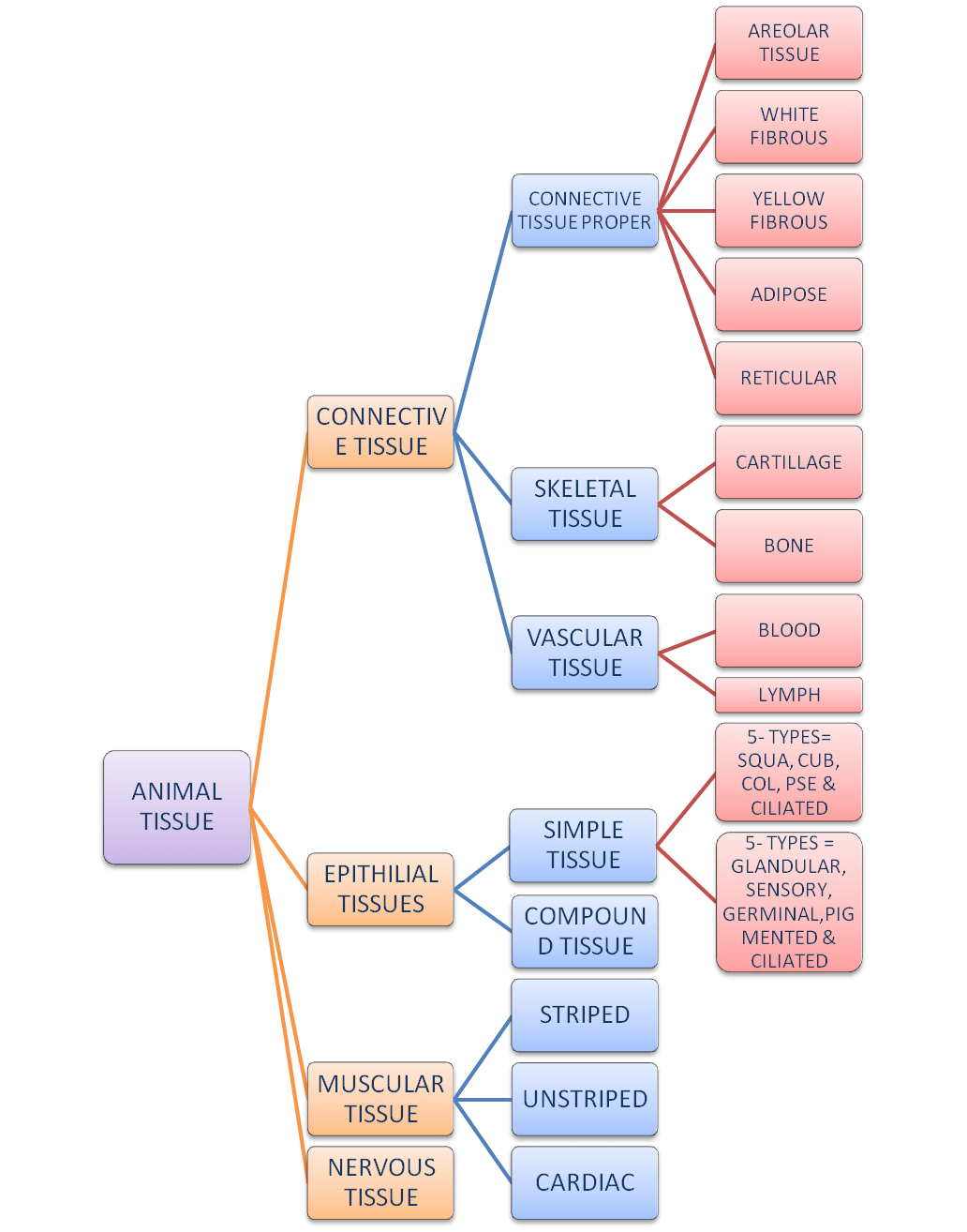
- EPITHELIAL TISSUES
This tissue has a free surface, which faces either a body fluid or the outside environment and thus provides a covering or a lining for some part of the body. The cells are compactly packed with little intercellular matrix. There are two types of epithelial tissues namely
- Simple epithelium– Simple epithelium is composed of a single layer of cells and functions as a lining for body cavities, ducts, and tubes. On the basis of shape of cell, it is of five types:
Squamous epithelium- Squamous epithelium is made of a single thin layer of flattened cells with irregular boundaries. They are found in the walls of blood vessels, air sacs of lungs, etc. and are involved in functions like forming a diffusion boundary and prevent underlying parts from injury.
Cuboidal epithelium-The Cuboidal epithelium is composed of a single layer of cube-like cells. This is commonly found in ducts of glands and tubular parts of nephrons in kidneys and its main functions are secretion and absorption.
Columnar epithelium- The columnar epithelium is composed of a single layer of tall and slender cells. Their nuclei are located at the base. Free surface may have microvilli. They are found in the lining of stomach and intestine and help in secretion and absorption.
Pseudostartified epithelium- it consists of two types of cells, long pillar like and small triangular. It is found in lining of trachea, bronchi, etc.
Ciliated epithelium- If the columnar or Cuboidal cells bear cilia on their free surface they are called ciliated epithelium.
- Compound epithelium – Compound epithelium consists of two or more cell layers and has protective function as it does in our skin. Their main function is to provide protection against chemical and mechanical stresses. They cover the dry surface of the skin, the moist surface of buccal cavity, pharynx, inner lining of ducts of salivary glands and of pancreatic ducts
- Connective tissues
Connective tissues are most abundant and widely distributed in the body of complex animals. They are named connective tissues because of their special function of linking and supporting other tissues/organs of the body. They range from soft connective tissues to specialised types, which include cartilage, bone, adipose, and blood. In all connective tissues except blood, the cells secrete fibres of structural proteins called collagen or elastin. The fibres provide strength, elasticity and flexibility to the tissue. These cells also secrete modified polysaccharides, which accumulate between cells and fibres and act as matrix (ground substance).
- Connective tissue proper: it holds various tissues together in any organ. it is further divided into three types:
- Areolar – it is found below the skin, in kidney, testis/w and around the mussel. It helps to hold various tissues together. These cells have Mast cells that concerned with the allergy.
- White fibrous tissues – its matrix contain white fibres that make it inelastic. Sheets of these tissues are found to cover the bones, cartilage, kidney, etc. Bundles of these tissues called Tendons.
- Yellow fibrous connective tissues – these tissues are very elastic. Sheets of these tissues cover up the blood vessels. Bundles of these tissues called ligament.
- Adipose tissues – it is modified Areolar tissues in which the cell becomes very large and become oval. Cells are called adipocytes. It is found below the skin, around heart brain and below the eye balls. It helps in storage of food in the form of fats. It act as insulators and prevents loss of heat.
- Skeletal tissues: it is the hard connective tissues that forms supportive framework of body. It is of two types:
- Cartilage – this tissue is elastic, harder than connective tissue proper but softer than bone. Its elasticity is due to presence of protein chondrin. Spaces in its matrix are called lacunae, which contain cartilage forming cells called chondrocytes. It is of three type’s hyaline, fibrous and calcified. It form supportive framework of nose, pinna, etc.
- Bone – it is hardest connective tissues its matrix is so much hard due to presence of salts, such as calcium phosphate, etc. it help in locomotion. It protect internal delicate organ.
- Vascular tissues: it is the fluid tissues that form supportive frame work of body. It is of two types:
- Blood — Blood is a special connective tissue consisting of a fluid matrix, plasma, and formed elements.
- Lymph –The fluid present in the lymphatic system is called the lymph. Lymph is a colourless fluid containing specialised lymphocytes which are responsible for the immune responses of the body. Lymph is also an important carrier for nutrients, hormones, etc. Fats are absorbed through lymph in the lacteals present in the intestinal villi.
- Muscular tissues
Each muscle is made of many long, cylindrical fibres arranged in parallel arrays. These fibres are composed of numerous fine fibrils, called myofibrils. Muscle fibres contract (shorten) in response to stimulation, then relax and return to their uncontracted state in a Coordinate fashion. Their action moves the body to adjust to the changes in the environment and to maintain the positions of the various parts of the body. In general, muscles play an active role in all the movements of the body.
Muscles are of three types, skeletal, smooth, and cardiac.
- Skeletal muscle tissue is closely attached to skeletal bones. In a typical muscle such as the biceps, striated (striped) skeletal muscle fibres are bundled together in a parallel fashion. A sheath of tough connective tissue encloses several bundles of muscle fibres.
- Smooth muscles — The smooth muscle fibres taper at both ends and do not show striations. Cell junctions hold them together and they are bundled together in a connective tissue sheath. The wall of internal organs such as the blood vessels, stomach and intestine contains this type of muscle tissue. Smooth muscles are ‘involuntary’ as their functioning cannot be directly controlled. We usually are not able to make it contract merely by thinking about it as we can do with skeletal muscles.
- Cardiac muscle tissue is a contractile tissue present only in the heart. Cell junctions fuse the plasma membranes of cardiac muscle cells and make them stick together. Communication junctions (intercalated discs) at some fusion points allow the cells to contract as a unit, i.e., when one cell receives a signal to contract.
- Neural tissues
It is highly specialised tissue due to which animals are able to perceive and respond to stimuli. It is characterised by following properties:
- It contain specialised cell that called NEURON.
- These are capable of receiving and responding to internal and external stimulating.
- In this tissue Neurons are packed with in special cells called neuroglial.
NEURON Nerve cell is the unit of nervous system. A neuron is a microscopic structure composed of three major parts, namely, cell body, dendrites and axon. The cell body contains cytoplasm with typical cell organelles and certain granular bodies called Nissl’s granules. Short fibres which branch repeatedly and project out of the cell body also contain Nissl’s granules and are called dendrites.